


Radiologic Examination
Radiologic Examinations and other image testing usually takes place in a facility’s Radiology or Nuclear Medicine department can take place in the practitioner’s office or other exam room.
On this page...
- Plain Films (X-Ray)
- Fluoroscopy
- Computerized (Axial) Tomography (CT)
- Magnetic Resonance Imaging (MRI)
- Magnetic Resonance Angiography (MRA)
- Positron Emission Tomography (PET)
- Single Photon Emission Computed Tomography (SPECT)
- Radiologic Exam Using Contrast Media
- Ultrasonography/Mammography/3D mammography
- Virtual Scopes
- Sentinel Node Mapping
Plain Films/X-rays
A type of radiation used in the diagnosis and treatment of cancer and other diseases. In low doses, x-rays are used to diagnose diseases by making pictures of the inside of the body. In high doses, x-rays are used to treat cancer (https://www.cancer.gov)
An x-ray examination may require the taking of a number of pictures. This is referred to as an x-ray series and is summarized in one report. For example, a cervical spine series may be taken to investigate metastatic disease in a cancer patient.
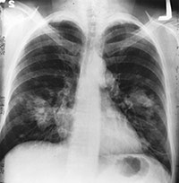
Figure 1. This is an x-ray image of a chest. Both sides of the lungs are visible with a growth on the left side of the lung, which could possibly be lung cancer.
Source: National Institutes of Health
Fluoroscopy
A technique for continuous or intermittent x-ray monitoring. X-ray images may be viewed directly without taking and developing x-ray photographs. This allows observation of certain dynamic body processes and is useful in certain surgical and diagnostic procedures. The radiologist moves the screen up and down the patient's body and observes what is happening within selected parts of the body. Fluoroscopy is especially useful for identifying the presence of restricted or blocked passages in the hollow organs of the body. For example, barium is swallowed and followed through the esophagus, stomach, and the upper intestinal tract.
Computerized (Axial) Tomography (CT)
A procedure that uses a computer linked to an x-ray machine to make a series of detailed pictures of areas inside the body. The pictures are taken from different angles and are used to create 3-dimensional (3-D) views of tissues and organs. A dye may be injected into a vein or swallowed to help the tissues and organs show up more clearly. A computerized tomography may be used to help diagnose disease, plan treatment, or find out how well treatment is working. CTs are also called computerized tomography and computerized axial tomography (CAT). (https://www.cancer.gov/)
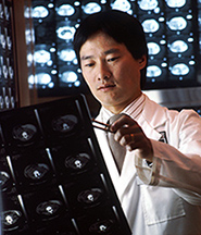
Figure 2. Radiologist reviewing computed tomography (CT) scans.
Source: Bill Branson (photographer), National Institutes of Health
Magnetic Resonance Imaging (MRI)
A procedure in which radio waves and a powerful magnet linked to a computer are used to create detailed pictures of areas inside the body. These pictures can show the difference between normal and diseased tissue. Magnetic resonance imaging makes better images of organs and soft tissue than other scanning techniques, such as computed tomography (CT) or x-ray. Magnetic resonance imaging is especially useful for imaging the brain, the spine, the soft tissue of joints, and the inside of bones. MRIs are also called NMRI, nuclear magnetic resonance imaging. (https://www.cancer.gov/)
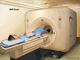
Figure 3. Magnetic resonance imaging (MRI) of the abdomen.
Source: Terese Winslow (illustrator), National Institutes of Health
Magnetic Resonance Angiography (MRA)
A procedure that uses radio waves and a powerful magnet linked to a computer to create detailed pictures of the blood vessels and blood flow inside the body. A dye may be injected into a vein to make the blood vessels and blood flow easier to see. Magnetic resonance angiography may be used to check for aneurysms (a bulge in the blood vessel wall), blockages in the arteries, blood clots, and other blood vessel problems. (https://www.cancer.gov/)
Positron Emission Tomography (PET) Scan
A procedure in which a small amount of radioactive glucose (sugar) is injected into a vein, and a scanner is used to make detailed, computerized pictures of areas inside the body where the glucose is taken up. Because cancer cells often take up more glucose than normal cells, the pictures can be used to find cancer cells in the body. Also called positron emission tomography scan. (https://www.cancer.gov/)
Single Photon Emission Computed Tomography (SPECT)
A special type of computed tomography (CT) scan in which a small amount of a radioactive drug is injected into a vein and a scanner is used to make detailed images of areas inside the body where the radioactive material is taken up by the cells. SPECT can give information about blood flow to tissues and chemical reactions (metabolism) in the body. (https://www.cancer.gov/)
Radiologic Examinations Using Contrast Media
Certain materials or gases can be injected into veins, arteries, lymphatics, or hollow cavities to obtain contrast with the surrounding tissues. A contrast medium is a radiopaque substance which obstructs the passage of x-rays so that the structures containing it appear white on the x-ray film, thus delineating abnormal pouches or growths and defining the contour of body structures on x-ray.
| Type of Radiologic Examination Using Contrast Media | Description of Radiologic Examination |
|---|---|
| Angiography | The radiographic study of the vascular system |
| Arteriography | X-ray examination of the arteries |
| Bronchogram | An x-ray of the bronchial system |
| Bronchography | The radiographic study of the bronchi of the lung |
| Cardiac Angiogram | X-ray showing the functions of the heart and large blood vessels |
| Cerebral Angiogram | X-ray of the vessels of the brain |
| Cholangiogram | X-ray of the extrahepatic bile ducts |
| Cholecystogram | X-ray of the gallbladder |
| Cholecystography | Radiologic study of the function of the gallbladder and bile ducts after an opaque medium has been introduced either orally or intravenously |
| Cystogram | X-ray of the urinary bladder by intravenous injection of a contrast medium |
| Infusion Nephrotomography | Radiologic visualization of the kidney by tomography after intravenous introduction of contrast medium |
| IVP (Intravenous Pyelography) | A succession of x-ray films of the urinary tract following the injection into a vein of an iodine-containing substance which is collected by the kidneys, passing into the ureters and subsequently the bladder, allowing the study of urinary tract function (also known as Intravenous Urography, IVU) |
| Lower GI Series or Barium Enema | X-ray studies, following rectal injection of the barium, of the large bowel |
| Lymphangiogram | X-ray study of the vessels of the lymphatic system |
| Myelography | Radiologic study of the spinal cord |
| Pneumocolon | X-ray examination of the colon in which air and barium are used as contrast materials; also known as double contrast enema and air contrast enema |
| Retrograde Urography; (Retrograde Pyelography) | Examination of the ureter and renal collection structures by means of installation of contrast material through a ureteral catheter passed through a cystoscope into the bladder and ureter |
| Salpingography | Radiologic study of the uterus and fallopian tubes |
| Sialography (Ptyalogram) | Radiologic study of the salivary ducts |
| Upper GI Series or Barium Swallow | X-ray studies, following ingestion of barium, of the pharynx, esophagus, stomach, duodenum, and small intestine |
Urogram (Pyelogram) |
X-ray of the kidney and ureter with emphasis on the pelvis of the kidney by intravenous injection of a contrast medium |
| Urography | Radiologic study of the urinary tract |
| Venography | X-ray examination of the veins |
Ultrasound
A procedure that uses high-energy sound waves to look at tissues and organs inside the body. The sound waves make echoes that form pictures of the tissues and organs on a computer screen (sonogram). Ultrasound may be used to help diagnose diseases, such as cancer. It may also be used during pregnancy to check the fetus (unborn baby) and during medical procedures, such as biopsies. Ultrasound may also be called ultrasonography. (https://www.cancer.gov/)
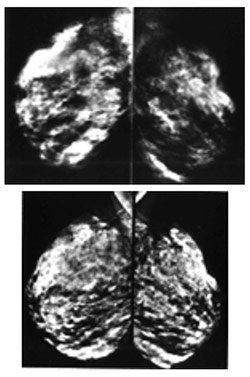
Figure 5. (Top) Breast images using conventional mammography. (Bottom) Digital mammography images of the same woman.
Source: Dr. Laurie Fajardo, Johns Hopkins Medical Institutions
The use of film or a computer to create a picture of the breast.
Digital Mammography
The use of a computer, rather than x-ray film, to create a picture of the breast. Mammography is available in both 2D and 3D forms.
A procedure that uses x-rays to take a series of pictures of the inside of the breast from many different angles. A computer is used to make 3-D pictures of the breast from these x-rays. 3-D mammography is used to check for breast cancer and other changes in the breast, such as abnormal lumps, cysts, or calcifications (calcium deposits). It may allow doctors to see breast tissue, including dense breast tissue, more clearly than with 2-D mammography. This may make breast tumors or other changes in the breast easier to find. 3-D mammography is also called 3-dimensional mammography and breast tomosynthesis. (https://www.cancer.gov/)
Virtual Colonoscopy
A method to examine the inside of the colon by taking a series of x-rays. A computer is used to make 2-dimensional (2-D) and 3-D pictures of the colon from these x-rays. The pictures can be saved, changed to give better viewing angles, and reviewed after the procedure, even years later. Virtual colonoscopy may also be called computed tomographic colonography, computed tomography colonography, CT colonography, and CTC. (https://www.cancer.gov/)
Sentinel Lymph Node Mapping
The use of dyes and radioactive substances to identify the first lymph node to which cancer is likely to spread from the primary tumor. Cancer cells may appear first in the sentinel node before spreading to other lymph nodes and other places in the body. (https://www.cancer.gov/)
Updated: December 28, 2023
Suggested Citation
SEER Training Modules: Radiologic Examination. U.S. National Institutes of Health, National Cancer Institute. Cited 12 February 2026. Available from: https://training.seer.cancer.gov.