


Diagnostic Surgery
There are many ways to detect or confirm a suspicion of the presence of a cancer. Microscopic examination of biopsy samples is the ideal way that a positive diagnosis of cancer can be made. This procedure involves physically removing all or part (tissue, cells, or fluid) of a suspected tumor and examining this material under a microscope. The purpose of a biopsy is to identify the histologic type of cancer and possibly define extent of disease.
Any organ in the body can be biopsied utilizing a variety of techniques. Some may require major surgery, while others may not even require local anesthesia. Types of biopsies include
- Bone marrow biopsy
- Colposcopic biopsy
- Core biopsy
- Excisional biopsy
- Endoscopic biopsy
- Fine needle aspiration biopsy
- Incisional biopsy
- Stereotactic biopsy
With the exception of excisional biopsy, biopsies typically leave gross tumor in the body.
The diagram that follows differentiates between an incisional biopsy and an excisional biopsy.
An incisional biopsy removes a small portion of the tumor to be analyzed by the pathologist to diagnosis the cancer. An incisional biopsy is considered a surgical diagnostic and staging procedure. The results of the biopsy if cancer is identified diagnoses the malignancy and depending on specifics about the specimen can contribute staging information.
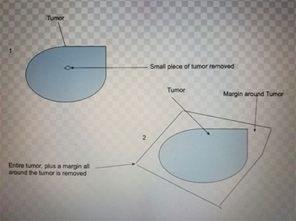
Figure 1. Picture depicting an incisional biopsy top left compared to an excisional biopsy bottom right of diagram.
An excisional biopsy of the primary site removes the entire tumor, plus a margin around the tumor. This procedure is considered a surgical procedure and treatment for the cancer. Often times the excisional biopsy may be the only surgical treatment the patient receives as the entire tumor is resected in this procedure.
Different types of endoscopes may be used in performing diagnostic and surgical procedures, these include
- Arthoscope
- Bronchoscope
- Colonoscope
- Colposcope
- Cystoscope
- Esophagoscope
- Gastroscope
- Laparoscope
- Laryngoscope
- Neuroendoscope
- Proctoscope
- Sigmoidoscope
The above scopes may be used for both surgical procedures and diagnostic procedures. The diagnostic procedures are discussed in detail in the “Abstracting the Medical Record” section of the training website.
The diagrams below depict a different endoscopic device. Endoscopic devices are named for the area they are used to examine.
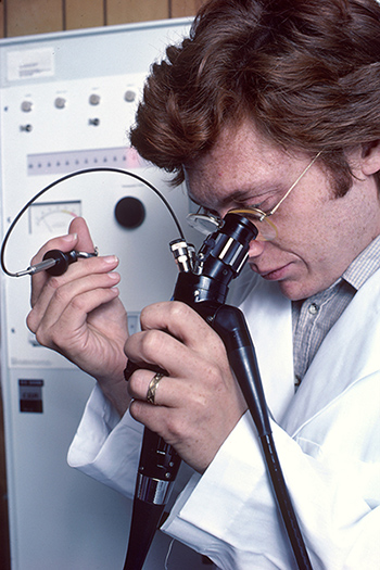
Figure 2. Endoscopy.
Source: Linda Bartlett (Photographer), National Cancer Institute.
Endoscopy: Figure 2 shows a Caucasian male physician using a remotely controlled endoscope. He is looking through a microscope-like eyepiece to monitor his actions while using small brushes and knives to take a biopsy.
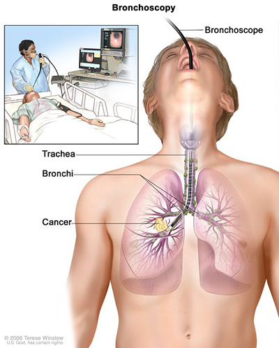
Figure 3. Bronchoscopy, with Cancer.
Source: Terese Winslow (Illustrator), National Cancer Institute.
Bronchoscopy, With Cancer - Bronchoscopy: Figure 3 shows a bronchoscope inserted through the mouth, trachea, and bronchus into the lung; lymph nodes along trachea and bronchi; and cancer in one lung. Inset shows patient lying on a table having a bronchoscopy.
A pathologist performs the gross and microscopic examination of the biopsied material. After careful evaluation, a benign or malignant diagnosis can usually be established. A written report prepared by the pathologist is sent to the physician that performed the diagnostic procedure. This report will also assist in the recommendations for a patient’s cancer treatment.
The procedures listed above are discussed in detail in the “Abstracting the Medical Record” section of the training website.
- Example: A bladder abnormality is suspected on physical examination due to presence of hematuria. The patient is referred to urology and a cystoscopy is done. During the cystoscopy a bladder tumor is identified which is suspicious for bladder cancer. A biopsy may be performed, or the entire area may be excised. The specimen taken is sent for pathological (microscopic) analysis of the abnormal tissue by the pathologists who can definitely confirm the presence of cancer.
Updated: December 21, 2023
Suggested Citation
SEER Training Modules: Diagnostic Surgery. U.S. National Institutes of Health, National Cancer Institute. Cited 09 February 2026. Available from: https://training.seer.cancer.gov.