




Tentorium
On this page...
The tentorium is a fold of the dura that separates the cerebellum from the cerebrum and divides the cranial cavity into supratentorial and infratentorial spaces.
- The supratentorium contains the cerebrum, ventricles (with cerebrospinal fluid in the figure below shown in blue), choroid plexus, hypothalamus, pineal gland, pituitary gland, and optic nerve.
- The infratentorium contains the cerebellum and brain stem.
Note: This distinction between the supratentorium (supratentorial) and the infratentorium (infratentorial) is relevant for coding EOD Primary Tumor and Summary Stage. See CNS Anatomy and ICD-O-3 for information on which subsites are supratentorial vs infratentorial (applicable for Brain primary sites only: C710-C719).
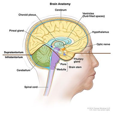
Brain Anatomy
Source: Terese Winslow (illustrator), National Institutes of Health
Brain
The Brain lies within the cranial cavity of the skull. It is described in terms of three main subdivisions
- Forebrain: Consists of the cerebrum (telencephalon) and the diencephalon
- The cerebrum is the part of the brain that receives and processes conscious sensation, generates thought, and controls conscious activity. It is the uppermost part of the brain and is divided into left and right hemispheres, which are joined by and communicated through the corpus callosum.
- Each cerebral hemisphere is divided into five lobes:
- Frontal lobe
- Parietal lobe
- Occipital lobe
- Temporal lobe
- Insula (Island of Reil)
- The cerebral hemispheres enclose the lateral ventricles (canals in the interior of the brain filled with cerebrospinal fluid). Each hemisphere consists of
- The cerebral cortex forms the outer layer. It contains the cell bodies of neurons often called the “gray mater” of the brain.
- The basal ganglia are deep within the hemispheres and are responsible for voluntary muscle movement. Basal ganglia are masses of gray mater
- Olfactory bulbs (rhinocephalon) lie on the ventral surface of each cerebral hemisphere and are associated with the sense of smell (cranial nerve I)
- The diencephalon or thalamencephalon connects the cerebral hemisphere with the midbrain and forms the walls of the third ventricle.
- The thalamus is an important relay center for sensory fibers on their way to the cerebral cortex. Crude sensation and pain may be registered here
- The hypothalamus contains the centers for the autonomic nervous system controlling sleep, water balance, temperature regulation, and secretions from the pituitary gland
- The epithalamus contains the pineal body (gland)
- Midbrain: Receives impulses from the eye and ear. It serves as the center for visual and auditory reflexes. It also connects the forebrain to the hindbrain. Cranial nerves III and IV stem from the midbrain.
- Hindbrain: Contains the pons, cerebellum, and the medulla oblongata. It surrounds the fourth ventricle.
- The pons bridges the cerebrum and the cerebellum. It contains nerve fibers which link the cerebral cortex with the medulla oblongata and the spinal cord.
- The nuclei of the cranial nerves V, VI, VII, and VIII are situated in the pons.
- The medulla oblongata controls breathing and heart rate. It consists of neurons which form the nuclei of the cranial nerves IX, X, XI, XII
- The cerebellum has centers which are concerned with balance and equilibrium. The function of the cerebellum is coordinating groups of muscles to work together smoothly.
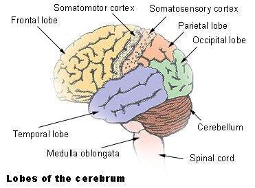
Lobes of the Cerebrum
Pituitary and Pineal Glands
These are endocrine organs that are located in the central nervous system.
- The pituitary gland (C751) secretes hormones, regulates and controls other hormone-secreting glands. It also regulates and controls many other body process, including reproduction.
- The craniopharyngeal duct (C752) serves as a developmental conduit for the formation of the pituitary gland. It is a short, temporary structure that links the front part of the developing pituitary (originating from the Rathke’s pouch) to the nasal cavity, allowing it to grow and obtain its mature form.
- The pineal gland (C753) produces and secretes melatonin which regulates the body’s circadian rhythm
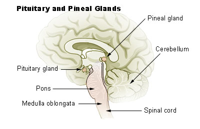
Lobes of the Cerebrum
Cranial Nerves
Twelve pairs of cranial nerves pass from the brain, through openings in the skull, to different areas of the head, neck, chest, and abdomen. They send information between the brain and the sense organs (the eyes, ears, nose, and tongue). They also send information to the muscles in the face, head, neck, and other organs in the body, including the larynx (voice box), heart, lungs, stomach, and intestines. The cranial nerves are part of the peripheral nervous system.
| Cranial Nerve # | Name of Cranial Nerves | Function | Type of Tumor |
|---|---|---|---|
| 1 | Olfactory (C722) | Cranial Nerve I, Olfactory Nerve I
|
Esthesioneuroblastoma |
| 2 | Optic (C723) | Cranial Nerve II, Optic Decussation, Optic Tract, Optic Nerve II
|
Optic nerve glioma |
| 3 | Oculomotor (C725) | Cranial Nerve III, Oculomotor III
|
Schwannoma |
| 4 | Trochlear (C725) | Cranial Nerve IV
|
Schwannoma |
| 5 | Trigeminal (C725) | Cranial Nerve V (three divisions)
|
Malignant peripheral nerve sheath tumor (MPNST) |
| 6 | Abducens (C725) | Cranial Nerve VI, Abducent Nerve
|
Schwannoma |
| 7 | Facial (C725) | Cranial Nerve VII
|
Schwannoma (rare) |
| 8 | Acoustic (C724) | Cranial Nerve VIII, Vestibulocochlear Nerve
|
Vestibular Schwannoma |
| 9 | Glossopharyngeal (C725) | Cranial Nerve IX
|
Glomus tumor |
| 10 | Vagus (C725) | Cranial Nerve X
|
Malignant peripheral nerve sheath tumor (MPNST), paraganglioma |
| 11 | Accessory (C725) | Cranial Nerve XI, Spinal Accessory Nerve
|
Schwannoma |
| 12 | Hypoglossal (C725) | Cranial Nerve XII
|
Schwannoma |
Spinal Cord
The spinal cord extends from the foramen magnum at the base of the skull to the level of the first lumbar vertebra. The cord is continuous with the medulla oblongata at the foramen magnum. Like the brain, the spinal cord is surrounded by bone, meninges, and cerebrospinal fluid.
The spinal cord is divided into 31 segments, with each segment giving rise to a pair of spinal nerves. At the distal end of the cord, many spinal nerves extend beyond the conus medullaris to form the cauda equina.
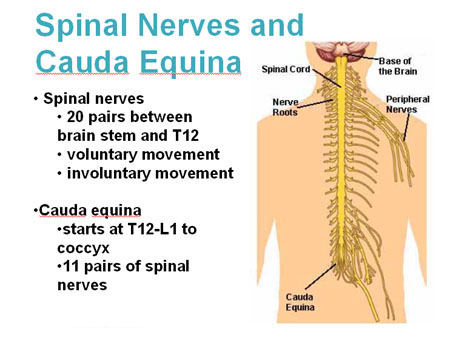
Spinal Nerves and Cauda Equina
Spinal Nerves
In certain areas of the spinal cord the anterior rami (branches) of the spinal nerves combine to form networks of nerve fibers called plexuses. These plexuses produce cerebrospinal fluid (CSF), which cushions the brain and spinal cord, provides nutrients, removes waste, and offers immunological protection. CSF is also known as a “shock absorber” for the brain and spinal cord.
- Thirty-one pairs of spinal nerves emerge laterally from the spinal cord. Consisting of, 8 cervical nerves, 12 thoracic nerves, 5 lumbar nerves, 5 sacral nerves, and 1 coccygeal nerve. Each pair of nerves corresponds to a segment of the cord and they are named accordingly.
- Each spinal nerve is connected to the spinal cord by a dorsal root and a ventral root. The cell bodies of the sensory neurons are in the dorsal root ganglion, but the motor neuron cell bodies are in the gray matter. The two roots join to form the spinal nerve just before the nerve leaves the vertebral column. Because all spinal nerves have both sensory and motor components, they are all mixed nerves.
- These nerve roots are located inside the spinal canal and covered by the spinal meninges.
| Plexus/Nerve | Number | Primary Site |
|---|---|---|
| Cervical | 8 pairs (C1-C8) | C470 |
| Thoracic | 12 pairs (T1-T12) | C473 |
| Lumbar | 5 pairs (L1-L5) | C721 |
| Sacral | 5 pairs (S1-S5) | C721 |
| Coccygeal | 1 pair (Co1) | C721 |
Lymph Nodes
There are no regional lymph nodes in the central nervous system.
Updated: September 23, 2026
Suggested Citation
SEER Training Modules: Tentorium. U.S. National Institutes of Health, National Cancer Institute. Cited 15 October 2026. Available from: https://training.seer.cancer.gov.