Code 7: Distant
- Tumor cells have broken away from the primary tumor, traveled to other part of the body, and started to grow at the new location
- Also referred to as remote, diffuse, disseminated, metastatic, or secondary disease
- No continuous trail of tumors cells between primary site and distant site (discontinuous)
- Common sites of distant metastases are liver, lung, brain, and bones
- These organs receive rich blood flow from all parts of the body and thus are a target for distant metastases
Types of distant routes of metastasis
- Hematogenous spread (via the blood)
- Lymphatic spread (via lymphatic system, including to distant lymph nodes)
- Implantation via fluids in bodily cavities
- Extension from the primary organ beyond adjacent tissue into the next organ
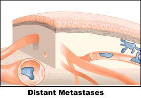
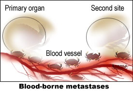
Source: Images adapted from illustrations by Brian Shellito of Scientific American, as printed in Cancer in Michigan, The Detroit News, Nov. 1-2, 1998.
- Hematogenous or blood-borne metastases
Invasion of blood vessels within the primary tumor allows escape of tumor cells or tumor emboli which are transported through the blood stream to another part of the body where it lodges in a capillary or arteriole. At that point the tumor penetrates the vessel wall and grows into the
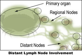
Source: Adapted from an illustration by Brian Shellito of Scientific American, as printed in Cancer in Michigan, The Detroit News, Nov. 1-2, 1998.
- Lymph channels beyond the first (regional) drainage area to lymph nodes are identified as being remote (distant) from the primary site.
- Tumor cells can be filtered, trapped and begin to grow in any lymph nodes in the body.
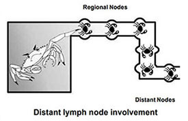
Source: Young JL Jr, Roffers SD, Ries LAG, Fritz AG, Hurlbut AA (eds). SEER Summary Staging Manual - 2000: Codes and Coding Instructions, National Cancer Institute, NIH Pub. No. 01-4969, Bethesda, MD, 2001.
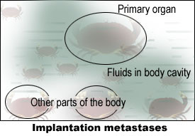
Source: Adapted from an illustration by Brian Shellito of Scientific American, as printed in Cancer in Michigan, The Detroit News, Nov. 1-2, 1998.
- Spread through fluids in a body cavity
- Example: malignant cells rupture the surface of the primary tumor and are released into the thoracic or peritoneal cavity. They float in the fluid and begin to grow on any tissue reached by the fluid.
- Implantation or seeding metastases
- Some tumors form large quantities of fluid called ascites that can be removed, but the fluid rapidly re-accumulates. However, the presence of fluid or ascites does not automatically indicate dissemination. There must be cytologic evidence of malignant cells.
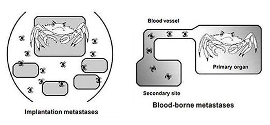
Source: Young JL Jr, Roffers SD, Ries LAG, Fritz AG, Hurlbut AA (eds). SEER Summary Staging Manual - 2000: Codes and Coding Instructions, National Cancer Institute, NIH Pub. No. 01-4969, Bethesda, MD, 2001.
Updated: November 5, 2024
Suggested Citation
SEER Training Modules: Code 7: Distant. U.S. National Institutes of Health, National Cancer Institute. Cited 28 February 2026. Available from: https://training.seer.cancer.gov.


