


Classification & Structure of Blood Vessels
Blood vessels are the channels or conduits through which blood is distributed to body tissues. The vessels make up two closed systems of tubes that begin and end at the heart. One system, the pulmonary vessels, transports blood from the right ventricle to the lungs and back to the left atrium. The other system, the systemic vessels, carries blood from the left ventricle to the tissues in all parts of the body and then returns the blood to the right atrium. Based on their structure and function, blood vessels are classified as either arteries, capillaries, or veins.
Arteries
Arteries carry blood away from the heart. Pulmonary arteries transport blood that has a low oxygen content from the right ventricle to the lungs. Systemic arteries transport oxygenated blood from the left ventricle to the body tissues. Blood is pumped from the ventricles into large elastic arteries that branch repeatedly into smaller and smaller arteries until the branching results in microscopic arteries called arterioles. The arterioles play a key role in regulating blood flow into the tissue capillaries. About 10 percent of the total blood volume is in the systemic arterial system at any given time.
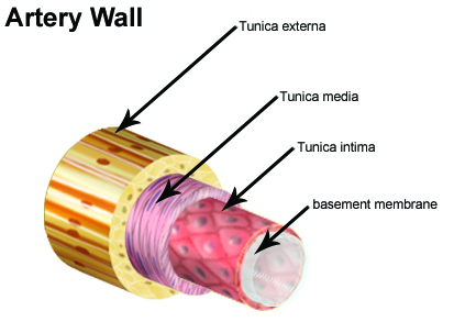
The wall of an artery consists of three layers. The innermost layer, the tunica intima (also called tunica interna), is simple squamous epithelium surrounded by a connective tissue basement membrane with elastic fibers. The middle layer, the tunica media, is primarily smooth muscle and is usually the thickest layer. It not only provides support for the vessel but also changes vessel diameter to regulate blood flow and blood pressure. The outermost layer, which attaches the vessel to the surrounding tissue, is the tunica externa or tunica adventitia. This layer is connective tissue with varying amounts of elastic and collagenous fibers. The connective tissue in this layer is quite dense where it is adjacent to the tunic media, but it changes to loose connective tissue near the periphery of the vessel.
Capillaries
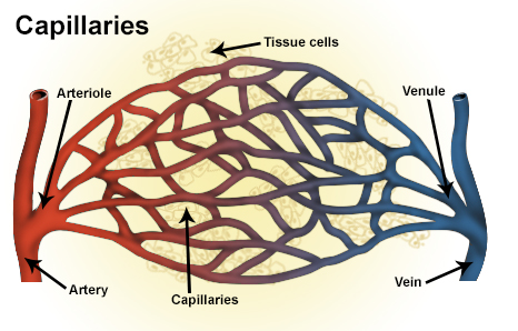
Capillaries, the smallest and most numerous of the blood vessels, form the connection between the vessels that carry blood away from the heart (arteries) and the vessels that return blood to the heart (veins). The primary function of capillaries is the exchange of materials between the blood and tissue cells.
Capillary distribution varies with the metabolic activity of body tissues. Tissues such as skeletal muscle, liver, and kidney have extensive capillary networks because they are metabolically active and require an abundant supply of oxygen and nutrients. Other tissues, such as connective tissue, have a less abundant supply of capillaries. The epidermis of the skin and the lens and cornea of the eye completely lack a capillary network. About 5 percent of the total blood volume is in the systemic capillaries at any given time. Another 10 percent is in the lungs.
Smooth muscle cells in the arterioles where they branch to form capillaries regulate blood flow from the arterioles into the capillaries.
Veins
Veins carry blood toward the heart. After blood passes through the capillaries, it enters the smallest veins, called venules. From the venules, it flows into progressively larger and larger veins until it reaches the heart. In the pulmonary circuit, the pulmonary veins transport blood from the lungs to the left atrium of the heart. This blood has a high oxygen content because it has just been oxygenated in the lungs. Systemic veins transport blood from the body tissue to the right atrium of the heart. This blood has a reduced oxygen content because the oxygen has been used for metabolic activities in the tissue cells.

The walls of veins have the same three layers as the arteries. Although all the layers are present, there is less smooth muscle and connective tissue. This makes the walls of veins thinner than those of arteries, which is related to the fact that blood in the veins has less pressure than in the arteries. Because the walls of the veins are thinner and less rigid than arteries, veins can hold more blood. Almost 70 percent of the total blood volume is in the veins at any given time. Medium and large veins have venous valves, similar to the semilunar valves associated with the heart, that help keep the blood flowing toward the heart. Venous valves are especially important in the arms and legs, where they prevent the backflow of blood in response to the pull of gravity.
Suggested Citation
SEER Training Modules: Classification & Structure of Blood Vessels. U.S. National Institutes of Health, National Cancer Institute. Cited 23 March 2026. Available from: https://training.seer.cancer.gov.