


Anatomy of the Liver & Gallbladder
Liver
The liver is the largest internal organ in the body. A primary liver cancer is uncommon; most liver tumors are metastatic. The liver is divided into two lobes, right (larger) and left.
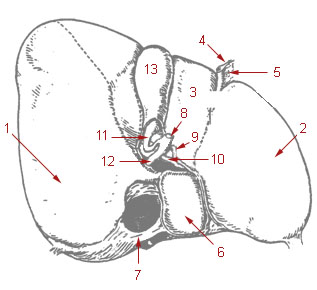
- Right lobe of liver
- Left lobe of liver
- Quadrate
- Round ligament
- Falciform
- Caudate lobe of liver
- Inferior vena cava
- Common bile duct
- Hepatic artery
- Portal vein
- Cystic duct
- Hepatic duct
- Gallbladder
Key words
- Porta hepatis: central region of liver which contains the hepatic artery, portal vein, lymphatic vessels, and extrahepatic bile ducts (referred to as portal area).
- Bile: fluid produced in the liver which aids in digestion.
Gallbladder
The gallbladder is a sac like organ; tumor extent is determined by invasion through the wall. The gallbladder lies under the liver and frequently (70 %) invades the liver by direct extension.
The gallbladder wall lacks the thick muscular layers of the bowel wall, but still has a mucosa, lamina propria, smooth muscle, and serosa (except on hepatic surface).
- Fundus: the fullest, most distal part of the gallbladder.
- Body: the area between the fundus and the neck of the bladder.
- Neck: tapers into cystic duct.
Ampulla of Vater
A dilated duct less than 1.5 cm in length below the pancreas which empties into duodenum.
Bile Ducts
Intrahepatic bile ducts in portal area between hepatic lobes, draining hepatic lobules.
Extrahepatic bile ducts:
- Common hepatic duct formed by junction of right and left hepatic bile ducts.
- Cystic duct connects common hepatic duct with gallbladder.
- Common bile duct formed by junction of cystic duct and common hepatic duct.
Suggested Citation
SEER Training Modules: Anatomy of the Liver & Gallbladder. U.S. National Institutes of Health, National Cancer Institute. Cited 10 February 2026. Available from: https://training.seer.cancer.gov.