


Anatomy of the Kidney & Ureter
Paired Organ: Yes.
Each kidney or ureter is considered a separate primary, unless bilateral involvement is stated to be metastatic from one side to the other (exception: bilateral Wilms tumor of the kidney).
The kidneys have two functional areas that are managed and staged independently, the kidney parenchyma and the renal pelvis.
The ureters are the tubes that carry urine from the renal pelvis to the bladder. They are staged the same way as the renal pelvis.
Anatomy of the Kidney and Ureter
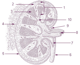
- Parenchyma
- Cortex
- Medulla
- Perirenal fat
- Capsule
- Ureter
- Pelvis of kidney
- Renal vessels
- Hilum
- Calyx
Key Words
Parenchyma
The solid part of the kidney, where the process of waste excretion takes place.
Cortex
The outer layer of the parenchyma consisting of connective tissue.
Glomeruli
Convoluted tubules where filtration is performed.
Medulla
Area of the kidney where filtration and concentration of wastes takes place, Henle's loops, pyramids of converging tubules.
Nephron
Basic functional unit of kidney.
Calyx (plural calyces)
Collecting area for urine within kidney before it is passed through to renal pelvis.
Capsule
Dense fibrous covering of kidney.
Pelvis
Central collecting system of kidney.
Hilum
Area of convergence of the renal collecting system, ureter, renal artery and vein.
Ureteropelvic junction
Point at which the renal pelvis becomes the ureter.
Gerota's fascia
Layer of connective tissue between the kidneys and the psoas muscles and the lumbar spine.
Perinephric fat
Layer of fat surrounding kidney outside of capsule.
Perihilar fat
Layer of fat in the area of the renal hilum.
Regional Lymph Nodes
| Site | Regional Lymph Nodes |
|---|---|
| Kidney | Para-aortic, paracaval, renal hilar nodes |
| Renal pelvis, ureter | Renal hilar, abdominal para-aortic, paracaval, common iliac, internal iliac, external iliac |
Suggested Citation
SEER Training Modules: Anatomy of the Kidney & Ureter. U.S. National Institutes of Health, National Cancer Institute. Cited 13 February 2026. Available from: https://training.seer.cancer.gov.