


Manipulative Procedures
There are a variety of diagnostic procedures which involve the direct viewing, feeling, hearing, and smelling of the body. Special instruments have been developed to provide the physician with a direct view the interior of the body and gross examination of any abnormalities.
An endoscope is an instrument for examining the interior of a canal or hollow viscus (any large interior organ). A variety of endoscopes have been developed for direct inspection of the interior of the stomach, larynx, colon, rectum, bladder, esophagus, and portions of the lung.
Manipulative procedures are usually performed in specialized surgical rooms. Some common endoscopic* examinations include
On this page...
- Bronchoscopy
- Colonoscopy
- Colposcopy
- Cystoscopy
- Esophagoscopy
- Gastroscopy
- Laparoscopy
- Laryngoscopy
- Mediastinoscopy
- Nasopharyngoscopy
- Ophthalmoscopy
- Otoscopy
- Panendoscopy
- Proctoscopy
- Sigmoidoscopy
- Thoracoscopy
Bronchoscopy
A procedure that uses a bronchoscope to examine the inside of the trachea, bronchi (air passages that lead to the lungs), and lungs. A bronchoscope is a thin, tube-like instrument with a light and a lens for viewing. It may also have a tool to remove tissue to be checked under a microscope for signs of disease. The bronchoscope is inserted through the nose or mouth. Bronchoscopy may be used to detect cancer or to perform some treatment procedures. (https://www.cancer.gov/)
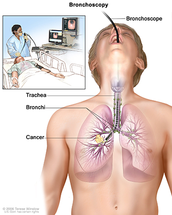
Figure 1. Bronchoscopy picture shows a bronchoscope inserted through the mouth, trachea, and bronchus into the lung; lymph nodes along trachea and bronchi; and cancer in one lung. A small inset picture shows patient lying on a table having a bronchoscopy.
Source: Terese Winslow (Illustrator), National Cancer Institute.
Colonoscopy
Examination of the inside of the colon using a colonoscope, inserted into the rectum. A colonoscope is a thin, tube-like instrument with a light and a lens for viewing. It may also have a tool to remove tissue to be checked under a microscope for signs of disease. (https://www.cancer.gov/)
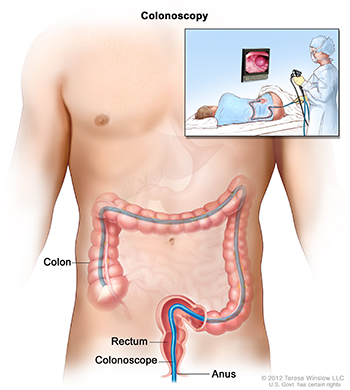
Figure 2. Colonoscopy picture shows a scope inserted through the anus and rectum and into the colon. A small inset picture shows patient on table having a colonoscopy.
Source: Terese Winslow (Illustrator), National Cancer Institute.
Colposcopy
A procedure in which a lighted, magnifying instrument called a colposcope is used to examine the cervix, vagina, and vulva. During colposcopy, an instrument called a speculum is inserted into the vagina to widen it so that the cervix can be seen more easily. A vinegar solution may be used to make abnormal tissue easier to see with the colposcope. Tissue samples may be taken using a spoon-shaped instrument called a curette and checked under a microscope for signs of disease. Colposcopy may be used to check for cancers of the cervix, vagina, and vulva, and changes that may lead to cancer. (https://www.cancer.gov/)
Cystoscopy
Examination of the bladder and urethra using a cystoscope, inserted into the urethra. A cystoscope is a thin, tube-like instrument with a light and a lens for viewing. It may also have a tool to remove tissue to be checked under a microscope for signs of disease. (https://www.cancer.gov/)
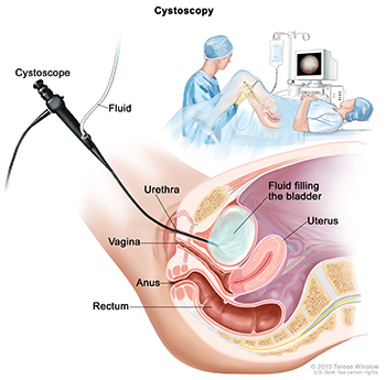
Figure 3. Cystoscopy picture shows a side view of the lower pelvis containing the bladder, uterus, and rectum. Also shown are the vagina and anus. The flexible tube of a cystoscope (a thin, tube-like instrument with a light and a lens for viewing) is shown passing through the urethra and into the bladder. Fluid is used to fill the bladder. A small inset picture shows a woman lying on an examination table with her knees bent and legs apart. She is covered by a drape. The doctor looks at an image of the inner wall of the bladder on a computer monitor.
Source: Terese Winslow (Illustrator), National Cancer Institute.
Esophagoscopy
Examination of the esophagus using an esophagoscope. An esophagoscope is a thin, tube-like instrument with a light and a lens for viewing. It may also have a tool to remove tissue to be checked under a microscope for signs of disease. (https://www.cancer.gov/)
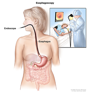
Figure 4. Esophagoscopy picture shows a scope inserted through the mouth and into the esophagus. A small inset picture shows patient on table having an esophagoscopy.
Source: Terese Winslow (Illustrator), National Cancer Institute.
Gastroscopy
Examination of the inside of the stomach using a gastroscope passed through the mouth and esophagus. A gastroscope is a thin, tube-like instrument with a light and a lens for viewing. It may also have a tool to remove tissue to be checked under a microscope for signs of disease. Also called upper endoscopy. (https://www.cancer.gov/)
Laparoscopy
A procedure that uses a laparoscope, inserted through the abdominal wall, to examine the inside of the abdomen. A laparoscope is a thin, tube-like instrument with a light and a lens for viewing. It may also have a tool to remove tissue to be checked under a microscope for signs of disease. https://www.cancer.gov/)
Laryngoscopy
Examination of the larynx (voice box) with a mirror (indirect laryngoscopy) or with a laryngoscope (direct laryngoscopy). (https://www.cancer.gov/)
Mediastinoscopy
A procedure in which a mediastinoscope is used to examine the organs in the area between the lungs and nearby lymph nodes. A mediastinoscope is a thin, tube-like instrument with a light and a lens for viewing. It may also have a tool to remove tissue to be checked under a microscope for signs of disease. The mediastinoscope is inserted into the chest through an incision above the breastbone. This procedure is usually done to get a tissue sample from the lymph nodes on the right side of the chest. https://www.cancer.gov/)
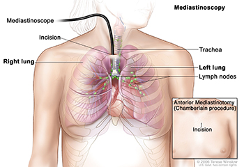
Figure 5. Mediastinoscopy picture shows a scope with light and lens inserted into the chest through an incision above the breastbone. Drawing shows right and left lungs, trachea, and lymph nodes. A small inset picture shows anterior mediastinotomy (Chamberlain procedure) with incision beside the breastbone.
Source: Terese Winslow (Illustrator), National Cancer Institute.
Nasopharyngoscopy
Examination of the nasopharynx, pharynx, and the pharyngeal end of the auditory tube by lighted telescopic endoscope.
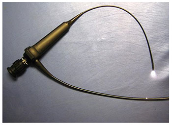
Figure 6. Adult fiberoptic nasopharyngoscope with 4-mm distal diameter, 2-way articulation, and video-recording capabilities.
Ophthalmoscopy
An exam that uses a magnifying lens and a light to check the fundus of the eye (back of the inside of the eye, including the retina and optic nerve). The pupils may be dilated (enlarged) with medicated eye drops so the doctor can see through the pupil to the back of the eye. Ophthalmoscopy may be used to check for eye problems, such as glaucoma, macular degeneration, eye cancer, optic nerve problems, or eye injury. Also called fundoscopy and funduscopy. (https://www.cancer.gov/)
Otoscopy
Otoscopy is a diagnostic procedure that examines the inner structures of the ear using a specialised device called the otoscope or auriscope. It is primarily performed to diagnose any abnormalities or conditions that affected the ear, particularly its middle part where structures responsible for hearing and balance are found. https://www.docdoc.com.sg/info/procedure/otoscopy/![]()
Panendoscopy
A cystoscopy that permits wide angle viewing of the urinary bladder.
Proctoscopy
A procedure that uses a proctoscope to look inside the anus and rectum. A proctoscope is a thin, tube-like instrument with a light and a lens for viewing. It may also have a tool to remove tissue to be checked under a microscope for signs of disease. (https://www.cancer.gov/)
Sigmoidoscopy
Examination of the lower colon using a sigmoidoscope, inserted into the rectum. A sigmoidoscope is a thin, tube-like instrument with a light and a lens for viewing. It may also have a tool to remove tissue to be checked under a microscope for signs of disease. Also called proctosigmoidoscopy. (https://www.cancer.gov/)
Thoracoscopy
Examination of the inside of the chest, using a thoracoscope. A thoracoscope is a thin, tube-like instrument with a light and a lens for viewing. It may also have a tool to remove tissue to be checked under a microscope for signs of disease. (https://www.cancer.gov/)
Updated: December 28, 2023
Suggested Citation
SEER Training Modules: Manipulative Procedures. U.S. National Institutes of Health, National Cancer Institute. Cited 18 July 2026. Available from: https://training.seer.cancer.gov.