


Colorectal Wall of Pathology
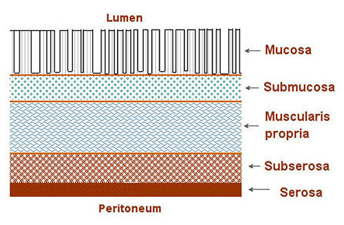
The colorectal wall is composed of 5 major layers.
| Layer | Explanation |
|---|---|
| Mucosal layer | Mucosal layer is the first of the 5 major layers. There are 4 sections of the mucosal layer This layer borders on the lumen, which is the interior surface of the colon “tube.” It contains no blood vessels or lymphatics. Also called surface epithelium |
| Mucosal layer | Second section of the Mucosal layer. A sheet of extracellular material, functions as a filtration barrier and a boundary involved in generating and maintaining tissue structures Also called Basement membrane |
| Mucosal layer | Third section of the Mucosal layer. Composed of areolar connective tissue, contains blood vessels, nerves, and, in some regions, glands. Once tumor has broken through the basement membrane into the lamina propria, it can spread by way of the lymphatic and blood vessels to other parts of the body Basement membrane/lamina propria are the dividing line between in situ and invasive tumors Also called the lamina propria |
| Mucosal layer | Fourth and final section of the Mucosal layer Thin layer of smooth muscle fibers. It is found in the wall of the digestive tract from the esophagus to the anal canal Also called the muscularis mucosae |
| Submucosa layer | The Submucosa layer is the second of the 5 major layers. It is a thick layer of either dense or areolar connective tissue. It contains blood vessels, lymphatic vessels, nerves, and in some regions, glands |
| Muscularis propria layer | The Muscularis propria layer is the third of the 5 major layers. It is a double layer of muscle tissue in most of the digestive tract; it constitutes the wall of the organ |
| Subserosa layer | The Subserosa layer is the fourth of the 5 major layers. This is the outermost layer covering most of the digestive tract, is a single layer of squamous, epithelial cells, part of the visceral peritoneum. Just inside the serosa (mesothelium) and sometimes considered part of the serosa, is a layer of connective tissue called the subserosa. The serosa and subserosa are present only in the peritonealized portions of the digestive tract. |
| Serosal layer | The Serosal layer is the last of the 5 major layers. This is the outer lining of organs and body cavities of the abdomen and chest, including the stomach. Includes the visceral peritoneum |
| Outside the serosa | This includes mesenteric fat, retroperitoneal fat or pericolic/perirectal fat |
Lymph Nodes
There are between 100 and 150 lymph nodes in the mesentery of the colon. Regional lymph nodes are the nodes along the colon, plus the nodes along the major arteries that supply blood to that particular colon segment.
| Segment | Regional Lymph Nodes |
|---|---|
| Cecum | Pericolic, anterior cecal, posterior cecal, ileocolic, right colic |
| Ascending colon | Pericolic, ileocolic, right colic, middle colic |
| Hepatic flexure | Pericolic, middle colic, right colic |
| Transverse colon | Pericolic, middle colic |
| Splenic flexure | Pericolic, middle colic, left colic, inferior mesenteric |
| Descending colon | Pericolic, left colic, inferior mesenteric, sigmoid |
| Sigmoid colon | Pericolic, inferior mesenteric, superior rectal, superior hemorrhoidal, sigmoidal, sigmoid mesenteric |
| Rectosigmoid | Perirectal, left colic, sigmoid mesenteric, sigmoidal, inferior mesenteric, superior rectal, superior hemorrhoidal, middle hemorrhoidal |
| Rectum | Perirectal, sigmoid mesenteric, inferior mesenteric, lateral sacral, presacral, internal iliac, sacral promontory (Gerota's) superior hemorrhoidal, inferior hemorrhoidal |
| Anus (included for reference) | Anorectal, inferior hemorrhoidal, inguinal (femoral) (deep and superficial), internal iliac, lateral sacral, mesorectal, obturator, perirectal and superior rectal (hemorrhoidal) (femoral |
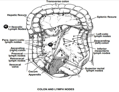
Sketch showing a colon and lymph nodes.
Source: Young JL Jr, Roffers SD, Ries LAG, Fritz AG, Hurlbut AA (eds). SEER Summary Staging Manual - 2000: Codes and Coding Instructions, National Cancer Institute, NIH Pub. No. No. 01-4969, Bethesda, MD, 2001.
Updated: June 24, 2025
Suggested Citation
SEER Training Modules: Colorectal Wall of Pathology. U.S. National Institutes of Health, National Cancer Institute. Cited 09 July 2026. Available from: https://training.seer.cancer.gov.