


Illustration of the Lung
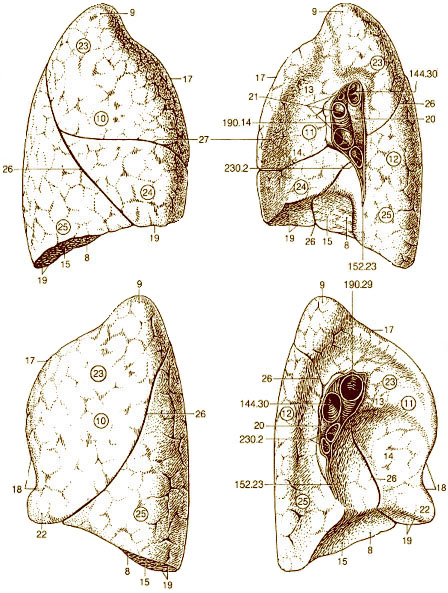
Illustration showing the anatomy of the lung.
Source: Pocket Atlas of Human Anatomy, 3rd edition. H. Feneis. Georg Thieme Verlag, New York. 1994.
- Base of lung
- Apex of lung
- Costal surface
Facing ribs - Medial surface
Facing mediastinum - Vertebral part
Facing vertebral column - Mediastinal part
- Cardiac impression
- Diaphragmatic surface
- Anterior border
- Cardiac notch
- Inferior border
- Hilum of lung
- Root of lung
- Lingula of left lung
- Superior lobe (upper lobe)
- Middle lobe (only on right)
- Inferior lobe (lower lobe)
- Oblique fissure
- Horizontal fissure of right lung
- 144.30 Main bronchus
- 152.23 Pulmonary ligament
- 190.14 Right pulmonary artery
- 190.29 Left pulmonary artery
- 230.2 Pulmonary veins
Suggested Citation
SEER Training Modules: Illustration of the Lung. U.S. National Institutes of Health, National Cancer Institute. Cited 24 March 2026. Available from: https://training.seer.cancer.gov.