


Anatomy
The lungs are part of the respiratory system. The respiratory system begins with the nose and ends the lungs. In between the nose and lungs are the nasopharynx, oropharynx, larynx, trachea and bronchi.
The thorax, which houses most of the organs of the respiratory system, plays a major role in respiration. The elliptical shape of the ribs and the angle of their attachment to the spine allows the thorax to expand during inspiration. Thus, the lungs provide a place where large amounts of oxygen can be taken from the air and quickly absorbed into the bloodstream. Conversely, large amounts of carbon dioxide are discharged into the lung when exhaled.
The rounded upper portion of each lung is called the apex; the base is the concave lower portion resting on the diaphragm. The hilus is the area on the medial surface through which the main bronchus, pulmonary artery, pulmonary vein, nerves, and lymph vessels enter and leave each lung. The left lung is partially divided by a fissure into an upper and lower lobe. Projecting from the lower portion of the left upper lobe is an area called the lingula (coded to left upper lobe). The right lung is divided by two fissures into three lobes (upper, middle, and lower). The left lung only has two lobes since the heart is in the way.
Visceral pleura or mesothelium is not part of the lung itself, but covers the outer surfaces of the lungs and adheres to them. Parietal pleura or mesothelium lines the thorax. The potential space between the visceral and parietal pleura is called the (inter)-pleural space or pleural cavity. The pleural space contains a lubricating pleural fluid which eliminates friction during the breathing process. This fluid drains to the mediastinal nodes. When the mediastinal nodes become inflamed or involved with a disease process such as a malignancy, a pleural effusion develops, causing breathing to become labored and painful.
The lungs provide a place where large amounts of oxygen can be loaded quickly into the blood and large amounts of carbon dioxide can be removed from it. The pulmonary artery from the right side of the heart branches into two arteries which carry deoxygenated blood to both lungs. Each artery continues dividing and subdividing within the lungs forming smaller and smaller vessels which end in capillaries which surround the alveolar sacs of the respiratory bronchioles. As the blood passes through the pulmonary capillaries, it absorbs oxygen and releases carbon dioxide. Then the newly oxygenated blood immediately returns through the venules (small veins) to the pulmonary veins which return to the left side of the heart.
The lung itself gets its nutrient supply from the bronchial arteries which branch off the aorta.
The lungs are the part of the body that make breathing (respiration) possible. Respiration can be divided into two distinct phases: external respiration and internal respiration
- External respiration involves both inspiration (inhaling) and expiration (exhaling) of air. Inspiration is the means by which oxygen is carried from the air into the lungs and then into the blood. Expiration is the means by which carbon dioxide is returned from the blood to the lungs and then expelled into the air.
- Internal respiration is concerned with the exchange of oxygen and carbon dioxide at the tissue level: the passage of oxygen from the blood to the tissues and the return of carbon dioxide from the tissues to the blood. First the oxygen is carried from the lungs by the red blood cells. Then carbon dioxide is carried by the red blood cells to the lungs. The utilization of oxygen and the elimination of carbon dioxide by the cells is called cellular respiration.
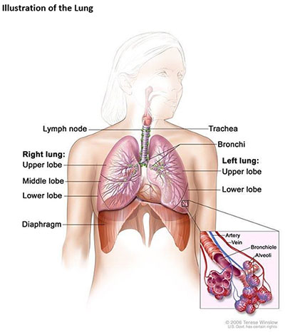
Source: Terese Winslow (illustrator), National Institutes of Health
Updated: April 8, 2026
Suggested Citation
SEER Training Modules: Anatomy. U.S. National Institutes of Health, National Cancer Institute. Cited 25 June 2026. Available from: https://training.seer.cancer.gov.