


Anatomy of the Female Pelvis
The upper two-thirds of the uterus is the body or corpus, which has its own diagnostic, staging and treatment guidelines. The lower third of the uterus comprises the cervix. The upper boundary of the cervix is the level of the internal os, a narrowing of the uterus that is also referred to as the isthmus. The internal os is the opening between the cervix and the corpus. The external os is the opening between the cervix and vagina. The cervix has no perimetrium (serosal covering).
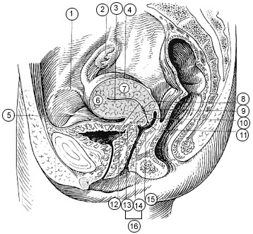
- Round ligament
- Uterus
- Uterine cavity
- Intestinal surface of Uterus
- Versical surface (toward bladder)
- Fundus of uterus
- Body of uterus
- Palmate folds of cervical canal
- Cervical canal
- Posterior lip
- Cervical os (external)
- Isthmus of uterus
- Supravaginal portion of cervix
- Vaginal portion of cervix
- Anterior lip
- Cervix
Within the cervix itself, the anatomy is subdivided into the endocervix and the exocervix or ectocervix. The upper two-thirds of the cervix (endocervix) contains columnar glandular epithelium. Adenosquamous carcinoma may arise here.
The lower third of the cervix (exocervix or ectocervix) is comprised of stratified squamous epithelium extending onto the lip of the cervix. The midpoint between the exocervix and the endocervix is the squamocolumnar junction, which is visible through a colposcope. The squamocolumnar junction is the most common primary site within the cervix.
The cervix projects into the vagina, and the circular trough formed at the upper end of the vagina around the cervix is the fornix. There are four fornices, two lateral, plus anterior and posterior. The lip or portio (portio vaginalis cervicis) is the portion of the cervix that extends into the vagina.
The Pouch of Douglas (cul-de-sac or rectovaginal septum) is the space between the rectum and the uterus. This is the lowest part of the abdominal cavity.
Suggested Citation
SEER Training Modules: Anatomy of the Female Pelvis. U.S. National Institutes of Health, National Cancer Institute. Cited 24 February 2026. Available from: https://training.seer.cancer.gov.