


Layers of the Skin
On this page...
The Epidermis
The epidermis is the outermost layer of the skin, and protects the body from the environment. The thickness of the epidermis varies in different types of skin; it is only .05 mm thick on the eyelids, and is 1.5 mm thick on the palms and the soles of the feet. The epidermis contains the melanocytes (the cells in which melanoma develops), the Langerhans' cells (involved in the immune system in the skin), Merkel cells and sensory nerves. The epidermis layer itself is made up of five sublayers that work together to continually rebuild the surface of the skin:
The Basal Cell Layer
The basal layer is the innermost layer of the epidermis, and contains small round cells called basal cells. The basal cells continually divide, and new cells constantly push older ones up toward the surface of the skin, where they are eventually shed. The basal cell layer is also known as the stratum germinativum due to the fact that it is constantly germinating (producing) new cells.
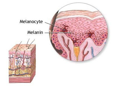
The basal cell layer contains cells called melanocytes. Melanocytes produce the skin coloring or pigment known as melanin, which gives skin its tan or brown color and helps protect the deeper layers of the skin from the harmful effects of the sun. Sun exposure causes melanocytes to increase production of melanin in order to protect the skin from damaging ultraviolet rays, producing a suntan. Patches of melanin in the skin cause birthmarks, freckles and age spots. Melanoma develops when melanocytes undergo malignant transformation.
Merkel cells, which are tactile cells of neuroectodermal origin, are also located in the basal layer of the epidermis.
The Squamous Cell Layer
The squamous cell layer is located above the basal layer, and is also known as the stratum spinosum or "spiny layer" due to the fact that the cells are held together with spiny projections. Within this layer are the basal cells that have been pushed upward, however these maturing cells are now called squamous cells, or keratinocytes. Keratinocytes produce keratin, a tough, protective protein that makes up the majority of the structure of the skin, hair, and nails.
The squamous cell layer is the thickest layer of the epidermis, and is involved in the transfer of certain substances in and out of the body. The squamous cell layer also contains cells called Langerhans cells. These cells attach themselves to antigens that invade damaged skin and alert the immune system to their presence.
The Stratum Granulosum & the Stratum Lucidum
The keratinocytes from the squamous layer are then pushed up through two thin epidermal layers called the stratum granulosum and the stratum lucidum. As these cells move further towards the surface of the skin, they get bigger and flatter and adhere together, and then eventually become dehydrated and die. This process results in the cells fusing together into layers of tough, durable material, which continue to migrate up to the surface of the skin.
The Stratum Corneum
The stratum corneum is the outermost layer of the epidermis, and is made up of 10 to 30 thin layers of continually shedding, dead keratinocytes. The stratum corneum is also known as the "horny layer," because its cells are toughened like an animal's horn. As the outermost cells age and wear down, they are replaced by new layers of strong, long-wearing cells. The stratum corneum is sloughed off continually as new cells take its place, but this shedding process slows down with age. Complete cell turnover occurs every 28 to 30 days in young adults, while the same process takes 45 to 50 days in elderly adults.
The Dermis
The dermis is located beneath the epidermis and is the thickest of the three layers of the skin (1.5 to 4 mm thick), making up approximately 90 percent of the thickness of the skin. The main functions of the dermis are to regulate temperature and to supply the epidermis with nutrient-saturated blood. Much of the body's water supply is stored within the dermis. This layer contains most of the skins' specialized cells and structures, including:
- Blood Vessels
The blood vessels supply nutrients and oxygen to the skin and take away cell waste and cell products. The blood vessels also transport the vitamin D produced in the skin back to the rest of the body. - Lymph Vessels
The lymph vessels bathe the tissues of the skin with lymph, a milky substance that contains the infection-fighting cells of the immune system. These cells work to destroy any infection or invading organisms as the lymph circulates to the lymph nodes. - Hair Follicles
The hair follicle is a tube-shaped sheath that surrounds the part of the hair that is under the skin and nourishes the hair. - Sweat Glands
The average person has about 3 million sweat glands. Sweat glands are classified according to two types:- Apocrine glands are specialized sweat glands that can be found only in the armpits and pubic region. These glands secrete a milky sweat that encourages the growth of the bacteria responsible for body odor.
- Eccrine glands are the true sweat glands. Found over the entire body, these glands regulate body temperature by bringing water via the pores to the surface of the skin, where it evaporates and reduces skin temperature. These glands can produce up to two liters of sweat an hour, however, they secrete mostly water, which doesn't encourage the growth of odor-producing bacteria.
- Sebaceous glands
Sebaceous, or oil, glands, are attached to hair follicles and can be found everywhere on the body except for the palms of the hands and the soles of the feet. These glands secrete oil that helps keep the skin smooth and supple. The oil also helps keep skin waterproof and protects against an overgrowth of bacteria and fungi on the skin. - Nerve Endings
The dermis layer also contains pain and touch receptors that transmit sensations of pain, itch, pressure and information regarding temperature to the brain for interpretation. If necessary, shivering (involuntary contraction and relaxation of muscles) is triggered, generating body heat. - Collagen and Elastin
The dermis is held together by a protein called collagen, made by fibroblasts. Fibroblasts are skin cells that give the skin its strength and resilience. Collagen is a tough, insoluble protein found throughout the body in the connective tissues that hold muscles and organs in place. In the skin, collagen supports the epidermis, lending it its durability. Elastin, a similar protein, is the substance that allows the skin to spring back into place when stretched and keeps the skin flexible.
The dermis layer is made up of two sublayers:
The Papillary Layer
The upper, papillary layer, contains a thin arrangement of collagen fibers. The papillary layer supplies nutrients to select layers of the epidermis and regulates temperature. Both of these functions are accomplished with a thin, extensive vascular system that operates similarly to other vascular systems in the body. Constriction and expansion control the amount of blood that flows through the skin and dictate whether body heat is dispelled when the skin is hot or conserved when it is cold.
The Reticular Layer
The lower, reticular layer, is thicker and made of thick collagen fibers that are arranged in parallel to the surface of the skin. The reticular layer is denser than the papillary dermis, and it strengthens the skin, providing structure and elasticity. It also supports other components of the skin, such as hair follicles, sweat glands, and sebaceous glands.
The Subcutis
The subcutis is the innermost layer of the skin, and consists of a network of fat and collagen cells. The subcutis is also known as the hypodermis or subcutaneous layer, and functions as both an insulator, conserving the body's heat, and as a shock-absorber, protecting the inner organs. It also stores fat as an energy reserve for the body. The blood vessels, nerves, lymph vessels, and hair follicles also cross through this layer. The thickness of the subcutis layer varies throughout the body and from person to person.
Suggested Citation
SEER Training Modules: Layers of the Skin. U.S. National Institutes of Health, National Cancer Institute. Cited 16 June 2026. Available from: https://training.seer.cancer.gov.