


Tumor Markers
Key Information
both for differential diagnosis and to monitor when tumor recurs
Alpha-fetoprotein:
A serum test used as a tumor marker for teratocarcinoma or embryonal carcinoma of the testis. Elevated alpha-fetoprotein levels are not found in other histologies of testicular cancer, although they may be found in patients with hepatocellular cancer. Note: Observe the date of an alpha-fetoprotein study carefully. For Collaborative Stage Site Specific Factor 1 record the highest value after orchiectomy and prior to other treatment. Alpha-fetoprotein is also used as a marker postoperatively to monitor residual tumor. Also called: aFP, AFP, alpha-fetoglobulin. Normal range: Adults: < 15 ng/ml.
Beta Subunit HCG (human chorionic gonadotropin):
A serum test used as a tumor marker for testicular carcinoma. Beta-HCG levels are never found in normal men. When the presence of β-HCG is detected in serum it always indicates a malignancy. Also called: β-HCG, beta-HCG, beta chain HCG. Note: Observe the date of the beta-HCG study carefully. For Collaborative Stage Site Specific Factor 2 record the highest value after orchiectomy and prior to other treatment.
Beta-HCG is also used as a marker postoperatively to monitor residual tumor and the effectiveness of therapy. In patients with testicular cancer who have had an orchiectomy, the presence of beta-HCG will confirm the patient has residual cancer that requires further treatment. However, when beta-HCG does not exist in the serum, the presence of active cancer cannot be excluded, especially in patients who have been previously treated. Normal range: 0 ng/ml.
Use of Tumor Markers for Specific Cell Types of Testicular Cancer
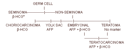
* "Pure" seminoma is incompatible with an elevated level of AFP.
Other Tumore Markers
LDH (Lactic Dehydrogenase): a blood chemistry study, usually part of a liver panel, useful in assessing liver and pulmonary disease. All tumors produce LDH.
Normal range: total LDH levels range from 48 to 115 IU/liter. There are five tissue-specific isoenzymes that can be identified and measured. The distribution of isoenzymes is as follows:
- LDH1: 18.1% to 29% of the total (heart, red blood cells and kidneys)
- LDH2: 29.4% to 37.5% of the total (heart, red blood cells and kidneys)
- LDH3: 18.8% to 26% of the total (lungs)
- LDH4: 9.2% to 16.5% of the total (liver and skeletal muscles)
- LDH5: 5.3% to 13.4% of the total (liver and skeletal muscles)
Notes: Not a screening test. Not diagnostic of testicular cancer. Elevated LDH is an indicator of possible tumor burden, such as metastatic involvement of liver or lung, and is elevated in 60% of patients with nonseminomatous germ cell tumors. LDH may be included in a Liver or Hepatic Panel/Profile, a Cardiac Panel, or general metabolic panel of tests. For Collaborative Stage Site Specific Factor 3 record the clinician's interpretation of the highest value prior to treatment.
PLAP (Placental Alkaline Phosphatase or PL-AP):
Differentiates the source of tumor among liver, bone, and germ cell origin; non-diagnostic by itself, it helps confirm malignancy in a small number of patients.
NSE (Neuron Specific Enolase):
Of secondary use in testicular neoplasms; elevated level indicates presence of tumor. Most useful in small cell carcinoma of the lung and neuroblastoma.
Go to the Tumor Markers page of the Diagnostic Tests module for more information.
Suggested Citation
SEER Training Modules: Tumor Markers. U.S. National Institutes of Health, National Cancer Institute. Cited 08 February 2026. Available from: https://training.seer.cancer.gov.