Anatomy
On this page...
Anatomy of the Female Breast
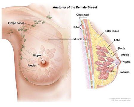
The breast is made up of lobes and ducts. Each breast has 15 to 20 sections called lobes, which are arranged in a circularfashion. The fat (subcutaneous adipose tissue) that covers the lobes gives the breast its size and shape. Each lobe has many smaller sections called lobules. Lobules end in dozens of tiny bulbs that can make milk. The lobes, lobules, and bulbs are linked by thin tubes called ducts. They are supported by and attached to the front of the chest wall on either side of the breastbone or sternum by ligaments. They rest on the major chest muscle, the pectoralis major.
The breast is responsive to a complex interplay of hormones that cause the tissue to develop, enlarge and produce milk. The three major hormones affecting the breast are estrogen, progesterone and prolactin, which cause glandular tissue in the breast and the uterus to change during the menstrual cycle and peripartum period.
The breast itself has no muscle tissue. A layer of fat surrounds the mammary glands. Mammary glands are located in the breast and produce milk for a baby after childbirth. Each gland contains lobules (or lobes) that produce the milk. Ductal carcinoma refers to cancer that starts in the mild ducts (mammary glands), while lobular carcinoma originates in the lobes (lobules).

Anatomy of the Female Breast.
Source: © 2011 Terese Winslow LLC.
Lymph Nodes
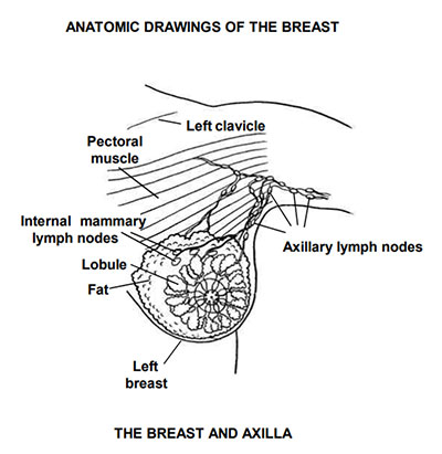
Each breast also has blood vessels and lymph vessels. The lymph vessels carry an almost colorless, watery liquid called lymph fluid. Lymph vessels carry lymph fluid between lymph nodes. Lymph nodes are small, bean-shaped structures found throughout the body. They filter lymph fluid and store white blood cells that help fight infection and disease. Groups of lymph nodes are found near the breast in the axilla (under the arm), above the collarbone, and in the chest.

Anatomic Drawings of the Breast.
Source: Summary Stage 1977.
The regional lymph nodes for breast are
- Axillary, NOS (ipsilateral)
- Axillary, NOS (ipsilateral)
- Level I (low-axilla)(low)(superficial), NOS [adjacent to tail of breast]
- Anterior (pectoral)
- Lateral (brachial)
- Posterior (subscapular)
- Level II (mid-axilla)(central), NOS
- Interpectoral (Rotter’s)
- Level III (high)(deep), NOS
- Apical (subclavian)
- Axillary vein
- Infraclavicular (subclavicular)(ipsilateral)
- Internal mammary (parasternal) (ipsilateral)
- Intramammary (ipsilateral)
- Supraclavicular (transverse) (ipsilateral)
Per the NCI Dictionary A sentinel lymph node is “defined as the first lymph node to which cancer cells are most likely to spread from a primary tumor. Sometimes, there can be more than one sentinel lymph node.” Sentinel lymph node biopsies are used in staging. If the sentinel lymph node biopsy is negative, then further surgery of the lymph nodes is not needed. If the sentinel lymph node biopsy is positive, then a lymph node dissection is done.
See Sentinel Lymph Node Biopsy for additional information on Sentinel Lymph Nodes.
See the current version of the SEER Program Coding Manual, Section VII: First course of therapy for information on the related lymph node data items and their coding instructions (Scope of Regional Lymph Nodes, Sentinel Lymph Nodes Examined, Sentinel Lymph Nodes Positive, Regional Nodes Examined, Regional Nodes Positive)
Breast Anatomy and ICD-O-3
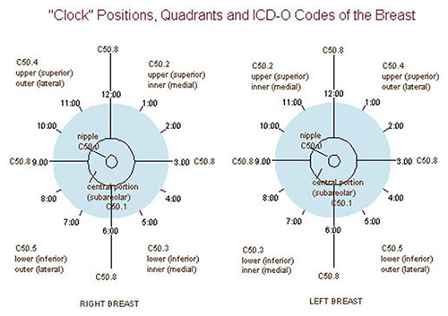
Each breast is divided into four quadrants-Upper inner, upper outer, lower inner, and lower outer, as well as a central portion that contains the areola and nipple. The location of the tumor in the breast is typically described as specific time on a clock.
- For Primary Sites, use ICD-O-3 (Topography section): ICD-O-3.1
or the Solid Tumor Rules
Priority order for coding primary site
- ICD-O
- SEER Program Manual (Including Coding Guidelines in Appendix C)
- Solid Tumor Rules (Breast)
| ICD-O-3 Codes | ICD-O-3 Preferred Term |
|---|---|
| C500 | Nipple |
| C501 | Central portion of breast |
| C502 | Upper inner quadrant of breast (UIQ) |
| C503 | Lower inner quadrant of breast (LIQ) |
| C504 | Upper outer quadrant of breast (UOQ) |
| C505 | Lower outer quadrant of breast (LOQ) |
| C506 | Axillary tail of breast |
| C508 | Overlapping lesion of breast (note: this is a single tumor which overlaps quadrants/subsite, or occurs directly at 3, 6, 9, 12 o’clock) |
| C509 | Breast, NOS |
See the current version of the SEER Program Coding Manual, Section IV: Description of this Neoplasm and Appendix C for complete coding instructions for primary site.
Updated: January 10, 2025
Suggested Citation
SEER Training Modules: Anatomy. U.S. National Institutes of Health, National Cancer Institute. Cited 28 February 2026. Available from: https://training.seer.cancer.gov.


