


Anatomy of the Esophagus
The esophagus is a muscular tube about ten inches (25 cm.) long, extending from the hypopharynx to the stomach. The esophagus lies posterior to the trachea and the heart and passes through the mediastinum and the hiatus, an opening in the diaphragm, in its descent from the thoracic to the abdominal cavity. The esophagus has no serosal layer; tissue around the esophagus is called adventitia.
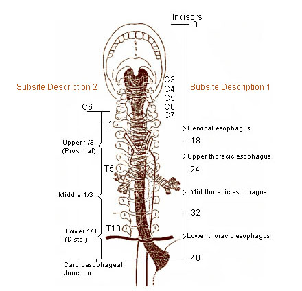
There are two sub site descriptions for the esophagus and they are not equivalent.
Sub Site Description 1
Cervical
- Cervical begins at the lower end of pharynx (level of 6th vertebra or lower border of cricoid cartilage) and extends to the thoracic inlet (suprasternal notch); 18 cm from incisors.
Thoracic
- Upper thoracic: from thoracic inlet to level of tracheal bifurcation; 18-23 cm.
- Mid thoracic: from tracheal bifuraction midway to gastroesophageal junction; 24-32 cm.
- Lower thoracic: from midway between tracheal bifurcation and gastroesophageal junction to GE junction, including abdominal esophagus; 32-40 cm.
Abdominal
- Considered part of lower thoracic esophagus; 32-40 cm.
Sub Site Description 2
- Upper third (10% of esophageal cancers)
- Middle third (40%)
- Lower third (50%)
The figure below illustrates the correlation between sub site descriptions of the esophagus.
Suggested Citation
SEER Training Modules: Anatomy of the Esophagus. U.S. National Institutes of Health, National Cancer Institute. Cited 16 July 2026. Available from: https://training.seer.cancer.gov.