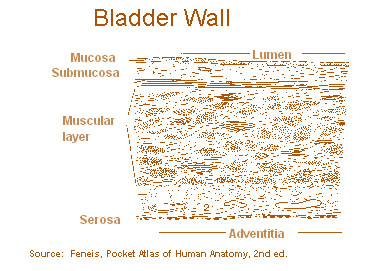
Layers of the Bladder Wall
The wall of the bladder wall has three principal tissue layers or coats:
- mucosa
- submucosa
- muscularis

Mucous membrane (mucosa)
Transitional epithelium; lines the bladder, ureters, and urethra
Epithelial layer
Contains no blood or lymphatic vesselsBasement membrane
Lies beneath the epithelial layer; single layer of cells separating the epithelial layer from the lamina propria; a sheet of extracellular material serving as a filtration barrier and supporting structure for the mucosal layerSubmucous coat (lamina propria)
Areolar connective tissue; interlaced with the muscular coat. This layer contains blood vessels, nerves, and in some regions, glands. A tumor, which has spread to this layer, can metastasize to the rest of the body via the lymphatics and blood vessels.
Muscular coat (muscularis propria)
Three layers: inner longitudinal, middle circular, and outer longitudinal
Serous coat (serosa)
A reflection of the peritoneum, which covers only the superior surface and the upper part of the lateral surfaces
Adventitia
In areas on bladder where there is no serosa, the connective tissue between organs merges
Perivesical fat
Layer of fat surrounding bladder outside of serosa/adventitia
Equivalent Terms for Layers of Bladder Wall
- epithelium
- urothelium
- mucosal surface
- transitional mucosa
- muscularis
- muscularis propria
- muscularis externa
- smooth muscle
- lamina propria
- suburothelial connective tissue
- subepithelial tissue
- stroma
- muscularis mucosa
Perivesical fat
- adventitia
- serosa
The most common sites for bladder tumors are the posterior and lateral walls. The superior wall is less frequently involved.
Key words:
Regional diathesis, field defect—terms which mean a tendency for the lining of the urinary tract to develop multiple tumors; a generalized deterioration of the urothelium from the renal pelvis into the urethra showing premalignant changes
