


Structure of the Heart
The human heart is a four-chambered muscular organ, shaped and sized roughly like a man's closed fist with two-thirds of the mass to the left of midline.
The heart is enclosed in a pericardial sac that is lined with the parietal layers of a serous membrane. The visceral layer of the serous membrane forms the epicardium.
Layers of the Heart Wall
Three layers of tissue form the heart wall. The outer layer of the heart wall is the epicardium, the middle layer is the myocardium, and the inner layer is the endocardium.
Chambers of the Heart
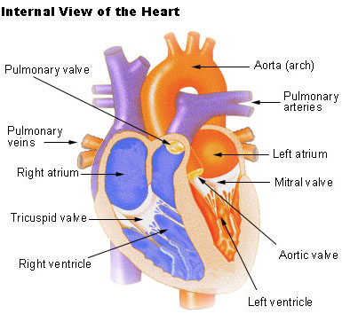
The internal cavity of the heart is divided into four chambers
- Right atrium
- Right ventricle
- Left atrium
- Left ventricle
The two atria are thin-walled chambers that receive blood from the veins. The two ventricles are thick-walled chambers that forcefully pump blood out of the heart. Differences in thickness of the heart chamber walls are due to variations in the amount of myocardium present, which reflects the amount of force each chamber is required to generate.
The right atrium receives deoxygenated blood from systemic veins; the left atrium receives oxygenated blood from the pulmonary veins.
Valves of the Heart
Pumps need a set of valves to keep the fluid flowing in one direction and the heart is no exception. The heart has two types of valves that keep the blood flowing in the correct direction. The valves between the atria and ventricles are called atrioventricular valves (also called cuspid valves), while those at the bases of the large vessels leaving the ventricles are called semilunar valves.
The right atrioventricular valve is the tricuspid valve. The left atrioventricular valve is the bicuspid, or mitral, valve. The valve between the right ventricle and pulmonary trunk is the pulmonary semilunar valve. The valve between the left ventricle and the aorta is the aortic semilunar valve.
When the ventricles contract, atrioventricular valves close to prevent blood from flowing back into the atria. When the ventricles relax, semilunar valves close to prevent blood from flowing back into the ventricles.
Pathway of Blood through the Heart
While it is convenient to describe the flow of blood through the right side of the heart and then through the left side, it is important to realize that both atria and ventricles contract at the same time. The heart works as two pumps, one on the right and one on the left, working simultaneously. Blood flows from the right atrium to the right ventricle, and then is pumped to the lungs to receive oxygen. From the lungs, the blood flows to the left atrium, then to the left ventricle. From there it is pumped to the systemic circulation.
Blood Supply to the Myocardium
The myocardium of the heart wall is a working muscle that needs a continuous supply of oxygen and nutrients to function efficiently. For this reason, cardiac muscle has an extensive network of blood vessels to bring oxygen to the contracting cells and to remove waste products.
The right and left coronary arteries, branches of the ascending aorta, supply blood to the walls of the myocardium. After blood passes through the capillaries in the myocardium, it enters a system of cardiac (coronary) veins. Most of the cardiac veins drain into the coronary sinus, which opens into the right atrium.
Suggested Citation
SEER Training Modules: Structure of the Heart. U.S. National Institutes of Health, National Cancer Institute. Cited 16 June 2026. Available from: https://training.seer.cancer.gov.