


Small & Large Intestine
Small Intestine
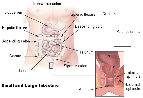
The small intestine extends from the pyloric sphincter to the ileocecal valve, where it empties into the large intestine. The small intestine finishes the process of digestion, absorbs the nutrients, and passes the residue on to the large intestine. The liver, gallbladder, and pancreas are accessory organs of the digestive system that are closely associated with the small intestine.
The small intestine is divided into the duodenum, jejunum, and ileum. The small intestine follows the general structure of the digestive tract in that the wall has a mucosa with simple columnar epithelium, submucosa, smooth muscle with inner circular and outer longitudinal layers, and serosa. The absorptive surface area of the small intestine is increased by plicae circulares, villi, and microvilli.
Exocrine cells in the mucosa of the small intestine secrete mucus, peptidase, sucrase, maltase, lactase, lipase, and enterokinase. Endocrine cells secrete cholecystokinin and secretin.
The most important factor for regulating secretions in the small intestine is the presence of chyme. This is largely a local reflex action in response to chemical and mechanical irritation from the chyme and in response to distention of the intestinal wall. This is a direct reflex action, thus the greater the amount of chyme, the greater the secretion.
Large Intestine
The large intestine is larger in diameter than the small intestine. It begins at the ileocecal junction, where the ileum enters the large intestine, and ends at the anus. The large intestine consists of the colon, rectum, and anal canal.
The wall of the large intestine has the same types of tissue that are found in other parts of the digestive tract but there are some distinguishing characteristics. The mucosa has a large number of goblet cells but does not have any villi. The longitudinal muscle layer, although present, is incomplete. The longitudinal muscle is limited to three distinct bands, called teniae coli, that run the entire length of the colon. Contraction of the teniae coli exerts pressure on the wall and creates a series of pouches, called haustra, along the colon. Epiploic appendages, pieces of fat-filled connective tissue, are attached to the outer surface of the colon.
Unlike the small intestine, the large intestine produces no digestive enzymes. Chemical digestion is completed in the small intestine before the chyme reaches the large intestine. Functions of the large intestine include the absorption of water and electrolytes and the elimination of feces.
Rectum and Anus
The rectum continues from the sigmoid colon to the anal canal and has a thick muscular layer. It follows the curvature of the sacrum and is firmly attached to it by connective tissue. The rectum ends about 5 cm below the tip of the coccyx, at the beginning of the anal canal.
The last 2 to 3 cm of the digestive tract is the anal canal, which continues from the rectum and opens to the outside at the anus. The mucosa of the rectum is folded to form longitudinal anal columns. The smooth muscle layer is thick and forms the internal anal sphincter at the superior end of the anal canal. This sphincter is under involuntary control. There is an external anal sphincter at the inferior end of the anal canal. This sphincter is composed of skeletal muscle and is under voluntary control.
Suggested Citation
SEER Training Modules: Small & Large Intestine. U.S. National Institutes of Health, National Cancer Institute. Cited 10 June 2026. Available from: https://training.seer.cancer.gov.