


Structure of Skeletal Muscle
A whole skeletal muscle is considered an organ of the muscular system. Each organ or muscle consists of skeletal muscle tissue, connective tissue, nerve tissue, and blood or vascular tissue.
Skeletal muscles vary considerably in size, shape, and arrangement of fibers. They range from extremely tiny strands such as the stapedium muscle of the middle ear to large masses such as the muscles of the thigh. Some skeletal muscles are broad in shape and some narrow. In some muscles the fibers are parallel to the long axis of the muscle; in some they converge to a narrow attachment; and in some they are oblique.
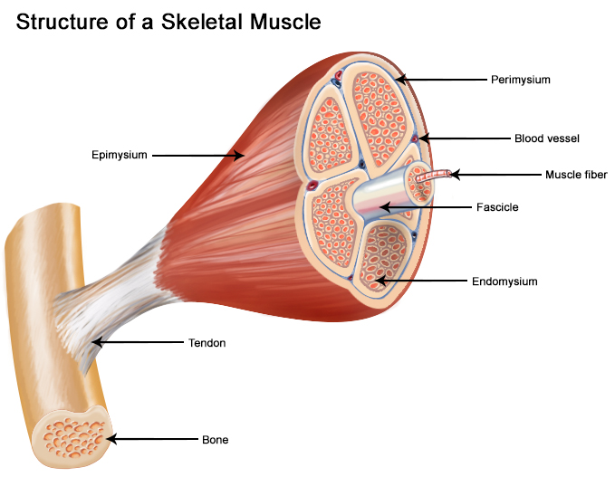
Each skeletal muscle fiber is a single cylindrical muscle cell. An individual skeletal muscle may be made up of hundreds, or even thousands, of muscle fibers bundled together and wrapped in a connective tissue covering. Each muscle is surrounded by a connective tissue sheath called the epimysium. Fascia, connective tissue outside the epimysium, surrounds and separates the muscles. Portions of the epimysium project inward to divide the muscle into compartments. Each compartment contains a bundle of muscle fibers. Each bundle of muscle fiber is called a fasciculus and is surrounded by a layer of connective tissue called the perimysium. Within the fasciculus, each individual muscle cell, called a muscle fiber, is surrounded by connective tissue called the endomysium.
Skeletal muscle cells (fibers), like other body cells, are soft and fragile. The connective tissue covering furnish support and protection for the delicate cells and allow them to withstand the forces of contraction. The coverings also provide pathways for the passage of blood vessels and nerves.
Commonly, the epimysium, perimysium, and endomysium extend beyond the fleshy part of the muscle, the belly or gaster, to form a thick ropelike tendon or a broad, flat sheet-like aponeurosis. The tendon and aponeurosis form indirect attachments from muscles to the periosteum of bones or to the connective tissue of other muscles. Typically a muscle spans a joint and is attached to bones by tendons at both ends. One of the bones remains relatively fixed or stable while the other end moves as a result of muscle contraction.
Skeletal muscles have an abundant supply of blood vessels and nerves. This is directly related to the primary function of skeletal muscle, contraction. Before a skeletal muscle fiber can contract, it has to receive an impulse from a nerve cell. Generally, an artery and at least one vein accompany each nerve that penetrates the epimysium of a skeletal muscle. Branches of the nerve and blood vessels follow the connective tissue components of the muscle of a nerve cell and with one or more minute blood vessels called capillaries.
Suggested Citation
SEER Training Modules: Structure of Skeletal Muscle. U.S. National Institutes of Health, National Cancer Institute. Cited 17 June 2026. Available from: https://training.seer.cancer.gov.